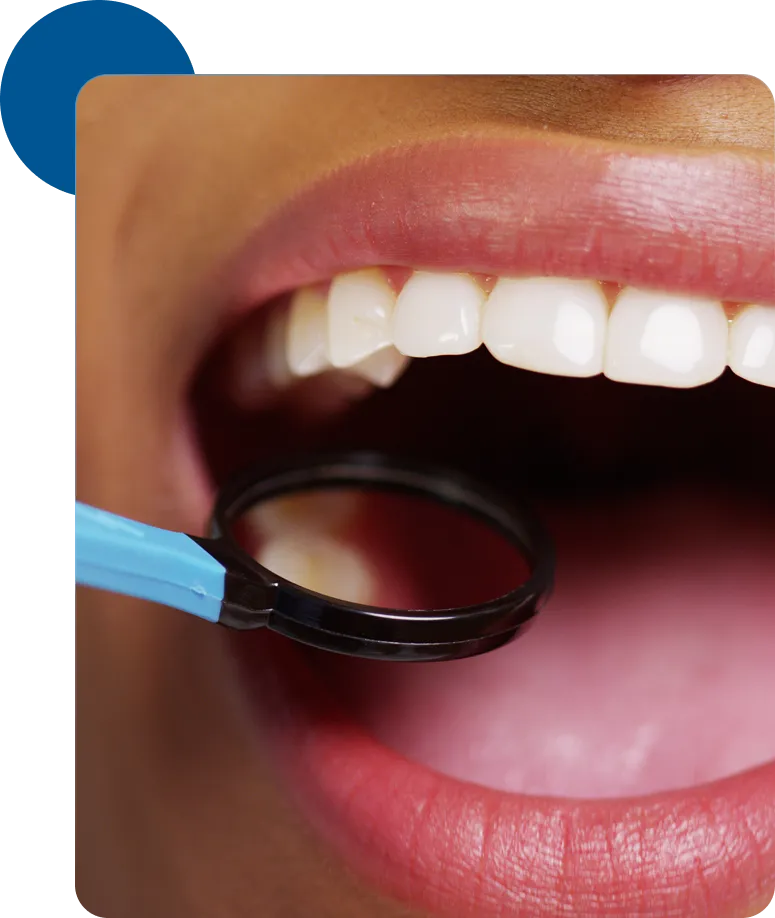
Intraoral Camera
Intraoral cameras used by dentists are quite small, approximately the size of an extra-thick
marker. Unlike the image of a large, unwieldy device that might come to mind, these cameras are
designed for comfortable use in the mouth.
Many dental conditions do not cause immediate pain or have obvious visual signs that are easily
visible without magnification. This can make it challenging for patients to comprehend the
specific issues that lead to a particular diagnosis. However, intraoral cameras are connected to
a TV screen or computer monitor located right beside the dental chair. This means that the
dentist can instantly display what the camera observes. Therefore, if you have concerns such as
swollen gums or a broken filling, you and the dentist can be on the same page regarding the
problem, even if you are not experiencing discomfort from it.
What Is An Intraoral Camera?
In simple terms, an intraoral camera is a small imaging device that takes photos of the
interior of your mouth. Unlike x-rays, which can reveal the internal structures of your
teeth and jaw, an intraoral camera provides highly detailed images of the exterior of your
teeth and gums.
Comparatively, dental x-rays have been in use since the discovery of x-rays in the 1890s,
while intraoral cameras gained widespread adoption only in the 1990s. Once a few dentists
began incorporating them into their practices, the dental community recognized their
potential to revolutionize patient care and diagnosis. At our practice, we view the use of
an intraoral camera as an essential part of our equipment, and we take pride in having this
powerful tool at our disposal to educate our patients and detect issues such as tooth decay,
damaged teeth, or restorations.
How An Intraoral Camera Works
The intraoral camera consists of a wand with a lens and a light on one end, with a cord leading
back to a computer on the other end. The camera's light is a crucial component. Even in well-lit
dental offices, it can be challenging to see into the darkest corners of your mouth. The
brightly lit images provided by the intraoral camera help us obtain a better view of your teeth
and gums. Many dental professionals find this method superior to traditional tools like a
headlamp and a mirror when it comes to detecting and diagnosing abnormalities.
One of the valuable features of an intraoral camera is its ability to capture excellent "before
and after" images. This is especially helpful when replacing an old metal filling (known as an
"amalgam" filling) with a tooth-colored filling (referred to as a "composite" filling). These
images make it easy to see the significant aesthetic improvement that white, natural-colored
fillings can offer.
Images taken with the intraoral camera primarily exist in the digital realm, although they can be
printed out if necessary. This digital format makes it simple to add photos to your digital
dental record. We can readily share images with other specialists involved in your care, dental
insurance companies, or dental lab technicians who are assisting in creating a custom tooth
restoration, such as a crown.
If you want to see what the dentist is observing, don't hesitate to ask us to show you using our
intraoral camera during your next appointment.